(1) 接受国家留学基金委(资助号:202106845011)和 CREST JPMJCR2105 项目的资助,于 2022 年 5 月 7 日至 2024 年 5 月 6 日在筑波大学计算光学组负责全场扫频光学相干层析成像研究,朱越作为第一负责人,设计与搭建光学系统、与医学院合作进行动物实验等研究工作。
(2) 国家自然科学基金青年基金项目,62005123 , 增强型全场结构光相干编码断层成像方法研究,2021/01/01-2023/12/31 , 在研,主持。
(3) 江苏省自然科学基金青年基金项目,BK20190455,结构光全场OCT超分辨成像与支持向量分类模型研究,2019/07-2022/06,在研,主持。
(4) 教育部中央高校基本科研业务费,自主科研项目, 30919011226,全场OCT无色散移相及肝组织实时成像研究,2019/01 - 2020/12,在研,主持。
(5) 南京理工大学科研启动经费,AE89991/076,基于偏振移相的快速全场光学相干层析系统研制,2018/04-2021/04,在研,主持。
(6) 美国国家科学基金,Shedding New Light on the Miracle of Life,1351981,2013.03至2019.03,424683美元,已结题。
近五年发表论文:
1. Yue Zhu, Yuan Zhou, and Zhenyan Guo, "Fractal-based aberration-corrected full-field OCT," Biomed. Opt. Express 14, 3775-3797 (2023)
2. 田浩颍, 汤丰锐, 高万荣, & 朱越. (2022). 动态散射光测量在全场光学相干层析技术中的应用. 中国激光, 49(5), 0507007.
3. Y. Zhu, W. Gao, Z. Guo, Y. Zhou, and Y. Zhou, Liver tissue classification of en face images by fractal dimensionbased support vector machine, Journal of biophotonics, 13(4), e201960154, January 2020
4. Y. Zhu, and W. Gao, Single-shot wavelength-independent phase-shifting method for full-field optical coherence tomography, Applied optics, 58(4), 806-813, February 2019
5. G. T. Smith, L. Li, Y. Zhu, and A. K. Bowden, Low-power, low-cost urinalysis system with integrated dipstick evaluation and microscopic analysis, Lab on a Chip, 18(14), 2111-2123, June 2018
国际会议:
1. Y. Zhu, Makita, S., & Yasuno, Y. (2024, March). Theoretical consideration of maximum numerically correctable defocus of point-scanning and full-field optical coherence tomography. In Optical Coherence Tomography and Coherence Domain Optical Methods in Biomedicine XXVIII (Vol. 12830, pp. 36-40). SPIE.
2. Y. Zhu, Zhenyan Guo and Yuan Zhou. DMD-based structured illumination full-field optical coherence tomography. Optical Coherence Tomography and Coherence Domain Optical Methods in Biomedicine XXVII. Vol. 12367. SPIE, January 2023.
3. Y. Zhu White-light structured illumination full-field optical coherence microscopy for resolution enhancement. Optics & Photonics Japan 2022, Utsunomiya, Japan, 2022.11.13-2022.11.16
4. Y. Zhu, W. Gao, and J. Chen, Achromatic phase-shifting method for isolated tissue imaging with video-rate FF-OCT, Proc. SPIE 11228, Optical Coherence Tomography and Coherence Domain Optical Methods in Biomedicine XXIV, 112282P, February 2020
5. Z. Guo, J. Wang, Y. Zhu, and R. Guo, Study on the calibration technique for multi-directional affine projection in optical computerized tomography, 9th International Symposium on Advanced Optical Manufacturing and Testing Technologies: Optical Test, Measurement Technology, and Equipment, International Society for Optics and Photonics, 1083908, January 2019
更多:
1. W. Gao, and Y. Zhu, Fractal analysis of en face tomographic images obtained with full field optical coherence tomography, Annalen der Physik, 529(3), 1600216, March 2017
2. Y. Zhu, W. Gao, Y. Zhou, Y. Guo, F. Guo, and Y. He, Rapid and high-resolution imaging of human liver specimens by full-field optical coherence tomography, Journal of biomedical optics, 20(11), 116010, November 2015
3. Y. Zhu, W. Gao, and Y. Guo, A method of improving imaging quality of full-field optical coherence tomography, Acta Optica Sinica 35(5), 0517001-0517006, May 2015
4. Y. Zhu, and W. Gao, High-resolution imaging of biological tissue with full-field optical coherence tomography, Proc.SPIE 9330, Three-Dimensional and Multidimensional Microscopy: Image Acquisition and Processing XXII, 93301H, March 2015
5. Y. Zhu, and W. Gao, Imaging of Liver Biopsy Cells with Full-field Optical Coherence Tomography, Science & Technology Review, 32(34), November 2014
6. Y. Zhu, and W. Gao, High-resolution full-field optical coherence tomography for biological tissue, Chinese J Lasers, 41(8), 0804002, August 2014
7. Y. Guo, W. Gao, and Y. Zhu, Compensation Interferometer Based Tandem Full-Field Optical Coherence Tomography System, Laser&Optoelectronics Progress, 54, 011101, December 2016
8. F. Yang, W.Gao, and Y. Zhu, Hilbert Transform in Full-Field Optical Coherence Tomography, Laser&Optoelectronics Progress, 53, 111102, July 2016
9. W. Gao, Y. Chen, C. Liu, Q. Zhang and Y. Zhu, FPGA-based Rapid Full Field Optical Coherence Tomography, Acta Photonica Sinica, 45(6), June 2016
10. X. Zhu, W. Gao, and Y. Zhu, Improved Kohler illumination-based full-field optical coherence tomography system, Acta Optica Sinica. Sin 34(5), 0511002, May 2014
11. X. Zhang, W. Gao, C. Chen, H. Bian and Y. Zhu, Extraction and compensation for depth-resolved phase error in spectral domain optical coherence tomography, Chinese J Lasers, 41(2), 0204002 February 2014
3. Y. Zhu, and W. Gao, Single-shot wavelength-independent phase-shifting method for full-field optical coherence tomography, Applied optics, 58(4), 806-813, February 2019
已授权的发明专利
(1) 朱越;高万荣;全场结构光相干编码断层成像装置及方法, 2020-5-1 中国, ZL 2020 1 0366785.X
(2) 高万荣; 朱越; 单次采集无色散移相全场光学相干层析成像装置及方法, 2017-5-27, 中国, ZL201710387615.8
(2) 高万荣; 郭英呈; 朱越; 基于补偿干涉仪的串联式全场光学层析成像装置及方法, 2016-9-12, 中国, ZL201610814446.7.
发明专利
(1) 朱越,宋文超,张学辉,梁超粤;一种全场光学时空相干编码动态体成像装置及方法,202211424510.2, 2022-11-15。
AM Lab, Augmented Microscopy Laboratory
Lab Vision
以光学相干层析显微术为手段,突破传统病理诊断的局限,发展术中实时非侵入无标记数字诊断的现代增强型显微术。
Research on optical coherence microscopy to break through the limitations of pathological diagnosis, developing the modern augmented microscopy for real-time, non-invasive, label-free digital diagnosis during surgery.
Academic Goal
My long-term academic goal is to establish an Augmented Microscopy Laboratory (AM Lab) to explore the potential of morphological and biomechanical characteristics to diagnose the feasibility of regaining vision in ophthalmology. In the upcoming five years, I will continuously focus on computational optical coherence microscopy to break through the limitations of pathological diagnosis, developing the modern augmented microscope for real-time, non-invasive, label-free digital diagnosis. My recent research projects cover the establishment of full-field optical coherence tomography with sub-pixel structured illumination to offer high-resolution and contrast-augmented imaging modalities.
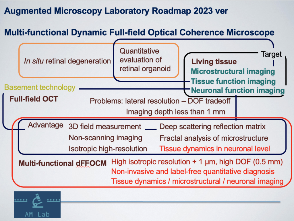
Roadmap
Contact
zhuyue[at]njust.edu.cn